|
|
|

|
See also: Crystallization of a protein by microseeding after establishing its phase diagramResearch by Yaakov M. Korkhin and Artem Evdokimov, Dept. of Structural Biology,
The Weizmann Institute of Science, Rehovot, Israel IntroductionThe microbatch technique is ideal for the rapid determination of the phase diagram of a protein: if the concentration of crystallizable protein is plotted against the concentration of a precipitant, microbatch results can be used to divide the space represented into several areas. This is shown schematically in the following figure. (i) At high concentrations of both protein and precipitant, the protein precipitates as
an amorphous material. It is often found that crystals grown in the metastable zone are better ordered and diffract better than crystals grown at higher concentrations. The microseeding approach adopted by Yaakov Korkhin and Artem Evdokimov described here includes a simple method of finding the metastable zone and introducing crystal seeds to it. Screening for initial conditionsA newly isolated bacterial enzyme was initially crystallized using Crystal Screen I by Hampton Research. PEG 3500 to 8000 was found to work well, with a range of buffers from pH 5.5 to 8.5. Ammonium acetate was initially thought to be an important additive. Attempts to crystallize with conventional vapor diffusionRepeated attempts were made to crystallize the protein with conventional hanging drop methods. The results were irreproducible, and crystals never diffracted beyond 3.5 A. The reason for this lack of success is not completely clear, but it is believed that pH fluctuation might have been responsible. Both ammonia and acetic acid are volatile, so they can travel from the well to the drop before equilibration is complete, causing the pH of the drop to vary. Use of IMPAX 1-5 to establish a phase diagramThe microbatch technique was used to establish a phase diagram for the protein with PEG 4000. For simplicity, ammonium acetate was not used, and it was later found that this material was not needed for successful crystallization. Dispensing took about four hours using automatic dispensing with IMPAX. Six protein concentrations were selected, and microbatch trials were dispensed at 24 different PEG concentrations for each protein concentration. Each set of 24 wells is represented by a horizontal line on the figure below.
Microbatch crystallization resultsTo illustrate the type of crystallization results obtained, we will show a typical line of 24 trials. The protein concentration giving the best crystals is shown. There is a small PEG increment between successive wells. These results are shown purely as an example, as the data are noisy and therefore not reproducible.
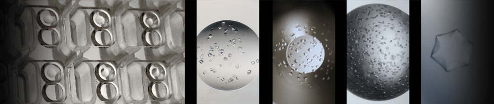
Starting from the lowest concentration of PEG, the first 12 wells were clear. The next well had small crystals. The next two contained a few large crystals. One of these is shown in Plate 1. The next well was clear (this result is ascribed to the extreme sensitivity of crystallization to dispensing errors). The remaining 8 wells had showers of crystals. Several good quality crystals were produced during the determination. It was believed that these grew in the metastable zone. Nuclei could have been formed in pockets with higher concentrations of protein and PEG, before concentration gradients were eliminated by diffusion. However, these crystals could not be grown reproducibly, and attempts to produce enough crystals for structure determination with microbatch were not successful. Generally no crystals grew for about two weeks, then poorly formed crystals such as those of Plate 2 grew very rapidly. Microseeding methodIt was therefore decided to try microseeding to control nucleation. The concentrations of all the solutions that were used were decided after careful study of the phase diagram. A series of microseeding solutions was produced as follows: 1. a well with a well formed crystal was selected Microseeding can be used with microbatch, but vapor diffusion was chosen for this project because it allowed the droplet concentration to be gently increased by about 10%. The four seeding solutions were used to seed sitting drop crystallization trials in Douglas Instruments' CrystalClear plates. 100 �l of solution was used in the reservoirs. This solution contained PEG at the same concentration as well 16. Next, buffer, protein and PEG were dispensed automatically into the sample wells of the CrystalClear plates using IMPAX. The PEG concentration of the droplets was the same as well 12. By dispensing droplets marginally below the reservoir concentration, the need for equilibration before seeding was avoided - the concentration was not so low that the nuclei dissolved. Finally 0.3 �l of each of the seeding solutions produced in step 7 above was added by hand to each sample well with a 5 �l Hamilton syringe. The plates were then sealed with Hampton tape in the usual way. The 10-3 dilution gave high quality diffracting crystals with great consistency. The typical size of crystals was 0.4 x 0.4 x 0.3 mm, and they diffracted using synchrotron radiation to 2 A. Typical crystals are shown in plate 3. Plate 4 shows the crystals produced by the normal vapor diffusion method without microseeding. Conditions are otherwise the same as those for plate 3. Note that seeding solutions are not stable and should be used immediately. When the seeding solution was used again after being stored for three days at 4�C, far fewer crystals were produced. DiscussionThe microbatch method made contributions to the project in two key areas: 1. Optimization: the optimum protein concentration and pH were determined. An
unnecessary additive was also eliminated with the minimum time and material. From the phase diagram the metastable zone was determined, which was used for microseeding. This gave a reliable supply of good quality crystals. Of course the phase diagram could have been determined by manual methods, but in practice few laboratories would have the resources, and few researchers the patience to use the fine coverage achieved here. The resulting fine resolution was crucial for clear understanding and the rational design of experiments. ReferencesFor a description of IMPAX and the microbatch method see the following three papers: N.E. Chayen, P.D. Shaw Stewart, D.L. Maeder, and D.M. Blow. An Automated System for Microbatch Protein Crystallization and Screening. J. Appl. Cryst. 23, pp 297-302 (1990). N.E. Chayen, P.D. Shaw Stewart, D.M. Blow. Microbatch Crystallization Under Oil - a New Technique Allowing Many Small-volume Crystallization Trials. J. Cryst. Growth, 122, pp 176-180 (1992). N.E. Chayen, P.D. Shaw Stewart, P. Baldock. New Developments of the IMPAX Small-Volume Automated Crystallization System. Acta Cryst. D. 50 pp 456-458 (1994). The following paper describes a rapid and accurate method of determining the precipitation point of a protein at a single protein concentration: P.D. Shaw Stewart, M. Khimasia. Predispensed Gradient Matrices - a New Rapid Method of Finding Crystallization Conditions. Acta Cryst. D. 50 pp 441-442 (1994). These two papers give a description of the determination and exploitation of the phase diagram of a protein, and also discuss questions of nucleation: E.E.G. Saridakis, P.D. Shaw Stewart, L.F. Lloyd, D.M. Blow. Phase Diagram and Dilution Experiments in the Crystallization of Carboxypeptidase G2. Acta Cryst. D. 50 pp 293-297 (1994). D.M. Blow, N.E. Chayen, L.F. Lloyd, E. Saridakis. Control of Nucleation of Protein Crystals. Protein Science, 3, pp 1638-1643 (1994) The last paper gives valuable insight into the control of nucleation in the case of lysozyme, which is often used as a model protein. N.E. Chayen, J. Radcliffe, D.M. Blow. The Control of Nucleation in the Crystallization of Lysozyme. Protein Science 2, pp 113-118 (1993).
Plate 1.
Plate 2.
Plate 3.
Plate 4.
|